There are three main sections in the Darkfield Microscopy course. It starts with creating a system that meets the needs of the practitioner. Those who have some experience can skip most of the Getting Started lesson.  So, the broader categories of what is covered are:

  1.  Configuration of a system that fits the budget and clinical or research requirements of the practice, assistance setting up the system for long distance collaboration, discussion of accessories and sampling techniques, and private tutoring that addresses the nuances of centering, focusing,  and skillful use of the microscope, camera, monitor, and record keeping system for documenting case histories.
  2.  Practice involving identification and correction of weaknesses and problems found; plus discussion of treatment options and interactions   between diet, herbs, supplements, and/or prescription medications.  Where indicated, toxins,  life styles, and environmental issues such as exposure to  electromagnetic interferences with physiological functioning are also addressed.
  3.  Mastering the use of natural remedies to perfect the plasma, erythrocytes, and white blood cells.

The course includes charts and written material as well as one-on-one sessions with Dr. Ingrid Naiman. There is a forum for discussion that is regulated by the level of enrollment.  There is also a secure area for uploading images and case histories that has a gallery for posting that has controlled access.

This course has prerequisites and requires a signed intellectual property agreement.

In some cases, additional charges will apply if the practitioner requests evaluations that go beyond what might be termed classroom as contrasted with clinical consultions.

Enrollment began in June 2022, and applications are currently being accepted.


Getting Started

Getting Started in Darkfield Microscopy


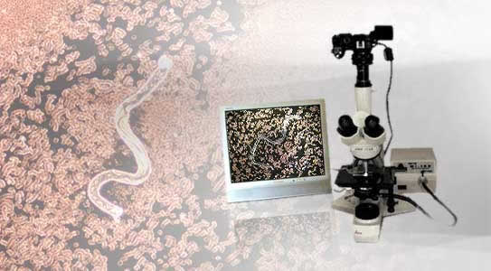
The first step in getting started with darkfield microscopy is to make sure that the system one is acquiring is configured to one's needs.  Once deciding on the model and camera, one needs a few basic supplies and usually some assistance in setting up the system and learning to use it.  Mistakes can be costly so it pays to have some help with the assembly and proper use of the equipment.

Next, of course, comes the sampling and there are some tips for preparing the slides and coverslips as well as how to take the best sample possible.  Then, the sample is placed on the microscope stage and explored.  Depending on one's background, what one does and does not actually observe can differ.  Being a medical doctor, hemotologist, pathologist, or microscopist does not necessarily prepare one for interpreting what is seen in darkfield.

The first step is to acquire a microscope so the Getting Started pdf will be sent when the student enrolls.  This will be followed by private tutorials.  The first real lesson will begin once the scope is in use.


Click to participate in this course

Rouleaux

Rouleaux
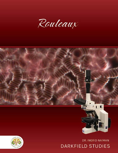
Rouleaux is often the first feature that students of darkfield microscopy identify.  The word, rouleaux, comes from the French term for a stack of coins and is actually the technical term used by microscopists.

There are numerous different causes for rouleaux as well as varying symptoms and risks.  In this lesson, the factors contributing to rouleaux as well as the corrections are covered in detail.  The course includes downloadable material, case histories, dietary suggestions, and herbal remedies for different underlying causes.

Course Date: 15 November 2022, 10 am PST


Click to participate in this course

Free Radicals

Image and text coming soon.

Course Date: To be announced

Click to participate in this course

Immunity

Image and text coming soon.

Course Date: To be announced


Click to participate in this course